
Technology HighLights, 2019
Technology HighLights, 2019
Complex 3D printing of human cells
Researchers have developed a method to 3D print cells to create human tissue such as ligaments and tendons, which could greatly improve a patient's recovery. In the future, their technique could lead to 3D printing of whole organs.
With today's technology, we can 3D-print sculptures, mechanical parts, prosthetics, and even guns and food. But a team of University of Utah biomedical engineers have developed a method to 3D-print cells to produce human tissue such as ligaments and tendons, a process that will greatly improve a patient's recovery. A person with a badly damaged ligament, tendon, or ruptured disk, for example, could simply have new replacement tissue printed and ultimately implanted in the damaged area.
"It will allow patients to receive replacement tissues without additional surgeries and without having to harvest tissue from other sites, which has its own source of problems," says assistant professor Robby Bowles, who worked with former biomedical engineering master's student, David Ede.
The 3D-printing method, which took two years to research, involves taking stem cells from the patient's own body fat and printing them on a layer of hydrogel to form a tendon or ligament, which would later grow in vitro in a culture before implantation. The process is extremely complicated, because that kind of connective tissue is made up of different cells in complex patterns. For example, cells that make up the tendon or ligament must then gradually shift to bone cells so the tissue can attach to the bone.
"This is a technique in a very controlled manner to create a pattern and organisations of cells that you couldn't create with previous technologies," Bowles says of the new process. "It allows us to very specifically put cells where we want them."
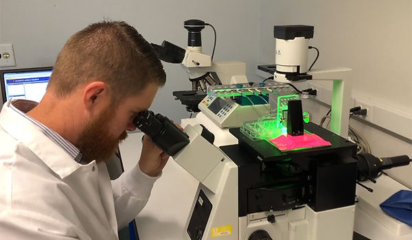
To do that, Bowles and his team worked with Salt Lake City-based company, Carterra, Inc., which develops microfluidic devices for medicine. Researchers used a 3D printer from Carterra typically used to print antibodies for cancer screening applications. But Bowles' team developed a special printhead for the printer that can lay down human cells in the controlled manner they require. To prove the concept, the team printed out genetically-modified cells that glow a fluorescent colour so they can visualise the final product, as shown in the image above.
Currently, replacement tissue for patients can be harvested from another part of the patient's body or sometimes from a cadaver, but they may be of poor quality. Spinal disks are complicated structures with bony interfaces that must be recreated to be successfully transplanted. This 3D-printing technique can solve those problems.
Bowles, a specialist in musculoskeletal research, said the technology is currently designed for creating ligaments, tendons and spinal discs, but "it literally could be used for any type of tissue engineering application," he says. It could even be applied to the 3D printing of whole organs, an idea researchers have been studying for years and that may be possible by 2025. Bowles also says the technology in the printhead could be adapted for any kind of 3D printer. His team's work is published in the Journal of Tissue Engineering, Part C: Methods.
Robotic surgery system arriving in 2019
A next-generation surgical robotic system has been developed in Cambridge, UK. It will be used on NHS patients for the first time next year.
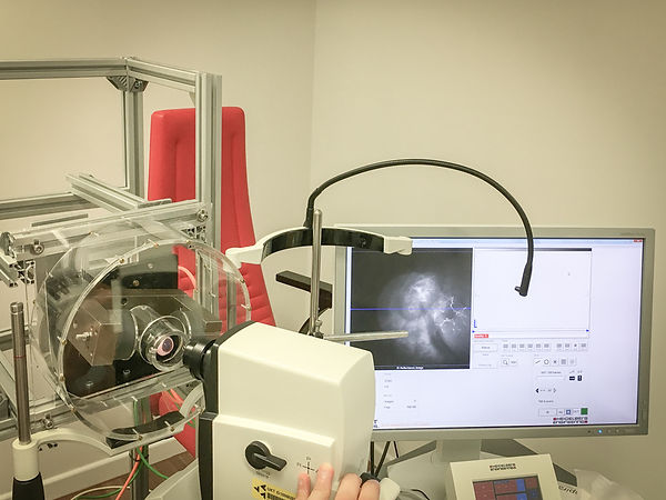
The Versius robot seen here was designed and built by CMR Surgical, formed in 2014, whose five founders brought with them global experience across the diverse disciplines of surgery, medical device development, start-ups and commercialisation. In June 2018, the company announced a record-breaking financing round that raised $100m in total, Europe’s largest private Series B medical device funding raise.
CMR's stated aim is "to make minimal access surgery available to all", by creating "a paradigm shift in robotic-assisted surgery". Minimal access surgery (sometimes known as keyhole or minimally invasive surgery) is an alternative to open surgery that was first pioneered around 40 years ago. For patients and healthcare providers alike, the benefits are numerous and compelling – reduced trauma, faster recovery and improved clinical outcomes. For example, the risk of infection from a robotically assisted hysterectomy is reduced by almost a factor of three compared with open surgery (from 6.5% to 2.2%).
There are an estimated six million open surgery procedures each year that could be performed using minimal access surgery. CMR Surgical believes that robotics opens up the potential for millions more people to benefit from laparoscopic surgery.
Versius, developed by CMR’s team of 100 scientists and engineers, is designed to meet the complex requirements of laparoscopic surgery and is intended to be used across a range of surgical specialities. Versatile and highly capable, its small and modular form factor and individually cart-mounted arms allow it to be moved effortlessly around the operating room, between operating suites and even hospitals. This combination of adaptability and portability allows hospital administrators to drive up utilisation of their robotics programme, improving hospital efficiency and clinical outcomes.
Versius mimics the dexterity and range of movement in the surgeon’s own hand and wrist, and is designed to be flexible enough to handle the majority of laparoscopic procedures. With 3D HD vision, easy-to-adopt instrument control and a choice of ergonomic working positions, the open surgeon console has been designed to reduce stress and fatigue; offering the potential to prolong a surgeon’s career. In addition, the versatility of the system and compelling commercial model allows healthcare providers to offer the benefits of robotic-assisted procedures in a cost-effective way.
"It takes around 80 hours to teach suturing with manual laparoscopic tools and some surgeons find it impossible to master," said Addenbrooke's Hospital surgeon Mark Slack, a co-founder of CMR Surgical. "By contrast, it takes half an hour to teach using Versius – this will enable many more surgeons to deliver the benefits of keyhole surgery."
The Versius machine is a rival to the American da Vinci system. CMR expects to introduce their new robotic system to hospitals within the next six months, beginning in the UK and Europe, then with international expansion shortly afterwards. The $4bn global market for surgical robots is predicted to increase five-fold to reach $20bn by 2024.
"Surgeons will remain in control, but as we develop the human-robot interface there may be simple parts of an operation, such as suturing or closing a wound that may be automated," said Dr Hachach-Haram, a member of the Royal College of Surgeons' Commission on the Future of Surgery.
In the coming decades, much greater advances may arrive. A large survey of experts in artificial intelligence, conducted last year, suggested that robotic surgery could be fully automated by 2053.
Nanobots propelled through eye tissue
Scientists developed specially coated nanometer-sized vehicles that can be actively moved through dense tissue like the vitreous of the eye. So far, the transport of nano-vehicles has only been demonstrated in model systems or biological fluids, but not in real tissue. The work was published in the journal Science Advances and constitutes one step further towards nanorobots becoming minimally-invasive tools for precisely delivering medicine to where it is needed.
Stuttgart – Researchers of the “Micro, Nano and Molecular Systems” Lab at the Max Planck Institute for Intelligent Systems in Stuttgart, together with an international team of scientists, developed propeller-shaped nanorobots that, for the first time, are able to drill through dense tissue as is prevalent in an eye.
They applied a non-stick coating to the nanopropellers, which are only 500 nm wide – exactly small enough to fit through the tight molecular matrix of the gel-like substance in the vitreous. The drills are 200 times smaller than the diameter of a human hair, even smaller than a bacterium´s width. Their shape and their slippery coating enable the nanopropellers to move relatively unhindered through an eye, without damaging the sensitive biological tissue around them. This is the first time scientists were able to steer nanorobots through dense tissue, as so far, it has only been demonstrated in model systems or biological fluids. The researchers´ vision is to one day load the nanopropellers with drugs or other therapeutic agents and steer them to a targeted area, where they can deliver the medication to where it is needed.
The molecule-matrix is like a tight mesh of double-sided adhesive tape
Targeted drug delivery inside dense biological tissue is very challenging, especially at these small scales: Firstly, it is the viscous consistency of the inside of the eyeball, the tight molecular matrix which a nanopropeller has to squeeze through. It acts as a barrier and prevents the penetration of larger structures. Secondly, even if the size-requirements are fulfilled, the chemical properties of the biopolymeric network in the eye would still result in the nanopropeller getting stuck in this mesh of molecules. Imagine a tiny cork-screw making its way through a web of double-sided adhesive tape.
And thirdly there is the challenge of precise actuation. This latter the scientists overcome by adding a magnetic material, like iron, when building the nanopropellers, which allows them to steer the drills with magnetic fields to the desired destination. The other obstacles the researchers overcome by making each nanopropeller not larger than 500 nm in size, and by applying a two layered non-stick coating. The first layer consists of molecules bound to the surface, while the second is a coating with liquid fluorocarbon. This dramatically decreases the adhesive force between the nanorobots and the surrounding tissue.
“For the coating we look to nature for inspiration”, the first author of the study Zhiguang Wu explains. He was a Humboldt Research Fellow at the MPI-IS and is now a postdoc at the California Institute of Technology. “In the second step, we applied a liquid layer found on the carnivorous pitcher plant, which has a slippery surface on the peristome to catch insects. It is like the Teflon coating of a frying pan. This slippery coating is crucial for the efficient propulsion of our robots inside the eye, as it minimizes the adhesion between the biological protein network in the vitreous and the surface of our nanorobots.”
“The principle of the propulsion of the nanorobots, their small size, as well as the slippery coating, will be useful, not only in the eye, but for the penetration of a variety of tissues in the human body”, says Tian Qiu, one of the corresponding authors of the paper, and who is a group leader in the “Micro, Nano and Molecular Systems” Lab at the MPI-IS.
Both Qiu and Wu are part of an international research team that worked on the publication with the title “A swarm of slippery micropropellers penetrates the vitreous body of the eye”. Also, the University of Stuttgart, the Max Planck Institute for Medical Research in Heidelberg, the Harbin Institute of Technology in China, Aarhus University in Denmark and the Eye Hospital of the University of Tübingen contributed to the groundbreaking work. It was at the eye hospital, where the researchers tested their nanopropellers in a dissected pig´s eye and where they observed the movement of the propellers with the help of optical coherence tomography, a clinical-approved imaging technique widely used in the diagnostics of eye diseases.
Across the eye towards the retina
With a small needle, the researchers injected tens of thousands of their bacteria-sized helical robots into the vitreous humour of the eye. With the help of a surrounding magnetic field that rotates the nanopropellers, they then swim towards the retina, where the swarm lands. Slippery nanorobots penetrate an eye. Being able to precisely control the swarm in real-time was what the researchers were aiming for.
But it doesn´t end here: the team is already working on one day using their nano-vehicles for targeted delivery applications. “That is our vision”, says Tian Qiu. “We want to be able to use our nanopropellers as tools in the minimally-invasive treatment of all kinds of diseases, where the problematic area is hard to reach and surrounded by dense tissue. Not too far in the future, we will be able to load them with drugs.“
For the Stuttgart scientists, this is not the first nanorobot they have developed. For several years now, they have been creating different types of nanorobots using a sophisticated 3D manufacturing process developed by the "Micro, Nano and Molecular Systems" research group led by Professor Peer Fischer. Billions of nanorobots can be made in only a few hours by vaporizing silicon dioxide and other materials, including iron, onto a silicon wafer under high vacuum while it turns. This is how the helix structure is created. See the video explaining the fabrication technique:
The full scientific paper can be found here: “A swarm of slippery micropropellers penetrates the vitreous body of the eye”, Zhiguang Wu, Jonas Troll, Hyeon-Ho Jeong, Qiang Wei, Marius Stang, Focke Ziemssen, Zegao Wang, Mingdong Dong, Sven Schnichels, Tian Qiu, Peer Fischer, Science Advances, 2018, DOI/10.1126/sciadv.aat4388

The 100,000 Genomes Project
The project has sequenced 100,000 genomes from around 85,000 people. Participants are NHS patients with a rare disease, plus their families, and patients with cancer.
The aim is to create a new genomic medicine service for the NHS – transforming the way people are cared for. Patients may be offered a diagnosis where there wasn’t one before. In time, there is the potential of new and more effective treatments.
The project will also enable new medical research. Combining genomic sequence data with medical records is a ground-breaking resource. Researchers will study how best to use genomics in healthcare and how best to interpret the data to help patients. The causes, diagnosis and treatment of disease will also be investigated. We also aim to kick-start a UK genomics industry. This is currently the largest national sequencing project of its kind in the world.
We have completed recruiting participants to the 100,000 Genomes Project. Results will be returned to the NHS throughout 2019.
Genetic testing, which may include whole genome sequencing where appropriate, is offered for certain medical conditions through the NHS Genomic Medicine Service.
GENOMICS ENGLAND AND THE 100,000 GENOMES PROJECT
Genomics England, with the consent of participants and the support of the public, is creating a lasting legacy for patients, the NHS and the UK economy, through the sequencing of 100,000 genomes.
In April 2003 one of the most significant scientific breakthroughs of modern times was announced. After years of painstaking research carried out by thousands of dedicated scientists across the world, the complete genetic code of a human being – their genome –could now be published.
The Human Genome Project, as this work was known, was the largest international collaboration ever undertaken in biology with British scientists leading the global race to read the human genome, which is made of DNA, letter by letter, a technique called sequencing. The UK has often led the world in scientific breakthroughs and DNA was no exception. Crick and Watson won the Nobel Prize for discovering the double helix structure of DNA and it was a British double Nobel Prize winning scientist, Fred Sanger, who discovered how to sequence it.
Now there is a real opportunity to turn the very important scientific discoveries about DNA and the way it works into a potentially life-saving reality for NHS patients across the country.
What is genomics?
Most of us have heard of genetics, the study of the way particular features or diseases are inherited through genes passed down from one generation to the next. But the more we learn about genes, the more we understand that the old idea of having a single gene for this, or a single gene for that, which determines your fate is not – except in the case of unusual inherited diseases - a good way of describing the complexity of genes. In fact, groups of genes work together and their activity is influenced by a huge variety of environmental and other factors. And we now know that the DNA between your genes is also very important.
You have a complete set of genes in almost every healthy cell in your body. One set of all these genes, (plus the DNA between the genes), is called a genome. Genomics is the study of the whole genome and how it works but has also come to have a broader meaning to include the way that the genome is interpreted and the technologies that have been developed to help do this.
Why this focus on genomics now?
When the first draft of the whole human genome was announced it was claimed that it would revolutionise medical treatment. It had taken 13 years and over £2 billion to laboriously read every letter of the human genetic code. It took such a long time because the DNA sequence of humans is very long – 3 billion letters – and because the sequencing machines available at the time were so slow and laborious. Now a human genome can be sequenced in a few days for less than £1000. It’s the leap in the speed and cost of technology that has opened up the potential of genomics and brought it within reach of mainstream healthcare.
But haven’t we already got a good understanding of genetics? One of the great surprises from the Human Genome Project was that there were only about 20,000 genes– about the same number as a starfish. The role of the remainder of a human’s genome – in fact a staggering 95 percent of it - was a mystery. Now we know that the remaining DNA is not irrelevant as was once thought but that much of it has a critically important role, influencing, regulating and controlling the rest. That’s why it’s
necessary to sequence the whole human genome (rather than just looking at the 20,000 genes currently used for diagnosis in medicine) if we are to really understand the role of genes in health and disease.
But people are very different, so studying only a small number of genomes would not be enough to give doctors and scientists a true picture of our genes and their relationship to disease. Another key point is that by itself, a genome can’t tell you very much. To make sense of it, it is essential to know much more about the person who donated it; details like their symptoms and when they first started, along with physiological measurements, such as heart rate or blood pressure (this sort of information is provided by clinicians and called phenotypic data). Another set of information which may be important in interpreting genomic data comes from their past medical records and would include such things as previous illnesses, medications and birth weight.
And this is where the NHS comes in. The way in which the NHS is able to link a whole lifetime of medical records with a person’s genome data and the fact it can do this on a large scale is unique. The richness of this data can help to understand disease and to tease apart the complex relationship between our genes, what happens to us in our lives and illness.
So what can genomics do? You can use it to predict how well a person will respond to a treatment or find one that will work best for them – so called personalised medicine. A good example in use already is whether or not a woman’s breast cancer is HER2 positive. If it is, Herceptin will be very effective for her but not for someone who doesn’t have HER2. You can also use genomics to test how well a cancer might respond to radiotherapy. For some that can mean far fewer radiotherapy sessions. Or use it to find the 30,000 people who currently use insulin for their Type 1 diabetes but would do better on simple tablets.
Genomics can be used to track infectious disease, precisely pinpointing the source and nature of the outbreak through looking at the whole genomes of bugs. The potential of genomics is
huge, leading to more precise diagnostics for earlier diagnosis, new medical devices, faster clinical trials, new drugs and treatments and potentially, in time, new cures.
The supersonic age of genomics has begun. And just as the NHS has been at the forefront of scientific breakthroughs before, we want the NHS to be at the forefront again, with its patients benefiting from all that genomics offers, becoming the first mainstream health service in the world to offer genomic medicine as part of routine care for NHS patients.
Human brain' supercomputer with 1 million processors switched on for first time
’ machine is capable of completing more than 200 SpiNNakerThe world’s largest neuromorphic supercomputer designed and built to work in the same way a human brain does has been fitted with its landmark one-millionth processor core and is being switched on for the first time. The newly formed million-processor-core ‘Spiking Neural Network Architecture’ or ‘million million actions per second, with each of its chips having 100 million transistors.
To reach this point it has taken £15million in funding, 20 years in conception and over 10 years in construction, with the initial build starting way back in 2006. The project was initially funded by the EPSRC and is now supported by the European Human Brain Project. It is being switched on for the first time on Friday, 2 November. The SpiNNaker machine, which was designed and built in The University of Manchester’s School of Computer Science, can model more biological neurons in real time than any other machine on the planet.
Biological neurons are basic brain cells present in the nervous system that communicate primarily by emitting ‘spikes’ of pure electro-chemical energy. Neuromorphic computing uses Instead it mimics the massively parallel communication architecture of the brain, sending billions of small amounts of information simultaneously to thousands of different destinations.large scale computer systems containing electronic circuits to mimic these spikes in a machine. SpiNNaker is unique because, unlike traditional computers, it doesn’t communicate by sending large amounts of information from point A to B via a standard network.
Steve Furber, Professor of Computer Engineering, who conceived the initial idea for such a computer, said: “SpiNNaker completely re-thinks the way conventional computers work. We’ve essentially created a machine that works more like a brain than a traditional computer, which is extremely exciting.
“The ultimate objective for the project has always been a million cores in a single computer for real time brain modelling applications, and we have now achieved it, which is fantastic.”
The computer’s creators eventually aim to model up to a billion biological neurons in real time and are now a step closer. To give an idea of scale, a mouse brain consists of around 100 million neurons and the human brain is 1000 times bigger than that. One billion neurons is 1% of the scale of the human brain, which consists of just under 100 billion brain cells, or neurons, which are all highly interconnected via approximately 1 quadrillion (that’s 1 with 15 zeros) synapses.
So, what is a million-core processor computer that mimics the way a brain works used for? One of its fundamental uses is to help neuroscientists better understand how our own brain works. It does this by running extremely large scale real-time simulations which simply aren’t possible on other machines.
For example, SpiNNaker has been used to simulate high-level real-time processing in a range of isolated brain networks. This includes an 80,000 neuron model of a segment of the cortex, the outer layer of the brain that receives and processes information from the senses.
It also has simulated a region of the brain called the Basal Ganglia - an area affected in Parkinson’s disease, meaning it has massive potential for neurological breakthroughs in science such as pharmaceutical testing. The power of SpiNNaker has even recently been harnessed to control a robot, the SpOmnibot. This robot uses the SpiNNaker system to interpret real-time visual information and navigate towards certain objects while ignoring others.
Prof Furber added: “neuroscientists can now use SpiNNaker to help unlock some of the secrets of how the human brain works by running unprecedentedly large scale simulations. It also works as real-time neural simulator that allows roboticists to design large scale neural networks into mobile robots so they can walk, talk and move with flexibility and low power.”


SpiNNaker completely re-thinks the way conventional computers work. We’ve essentially created a machine that works more like a brain than a traditional computer, which is extremely exciting.
Steve Furber, ICL Professor of Computer Engineering in the School of Computer Science
Computer logic meets cell biology, cell science getting upgrade
When manipulating the functions of cells, researchers need to take a multidisciplinary approach.
When Yvonne Chen published the first paper1 on a particular immune cell engineered to target either of two protein fragments on a cancer cell, several colleagues tried to discourage her from describing her creation in the unfamiliar language of computer logic. She did it anyway.
Adoptive T-Cell Therapy Attacks Leukemia - ACGT Scientist Dr. Yvonne Chen
Chen, a chemical and biomolecular engineer at the University of California, Los Angeles, had used synthetic proteins known as chimaeric antigen receptors (CARs) to modify the immune cell, a T lymphocyte, so that it could look for the two fragments — CD19 and CD20, examples of immune-system-stimulating molecules called antigens. This design meant that, if the cancer it was attacking underwent a mutation that rendered one antigen unrecognizable, the T cell could still use the other one to find and kill the cancer cell. Instead of referring to her creation using the existing biological terminology, as a bispecific cell, she called it an OR-gate CAR T cell, because of its ability to recognize one target or the other. “I actually received advice from multiple people saying, ‘You shouldn’t call it an OR-gate CAR, because people who work on T cells, who are either cell biologists or physicians, won’t understand what an OR gate means’,” she says. “But now everybody calls it an OR-gate CAR, because it makes sense.”
The concept of an OR gate is more familiar in computer science, where it refers to a logical operation that is triggered in the presence of either of two input options. But it describes in a few letters what the cell actually does, and distinguishes it from other types of bispecific CAR T cell, Chen says. Her OR-gate cell is nearly ready to begin clinical trials, and her lab is developing cells that mimic other logical functions, such as AND, which operates only if two inputs are positive, and NOT, which gives a negative output if the input is positive.
Merging fields
Terminology and concepts from computer science and engineering are becoming more common in biology labs as scientists re-engineer the activity of cells for specific applications. They are gaining unprecedented capabilities both from genetic-editing tools that have been around for a while, such as those that use viruses or proteins called zinc fingers, and from new CRISPR–Cas9 technology that allows more-targeted editing of DNA.
Scientists are creating vast collections of data by tweaking the transcription factors that copy DNA, one at a time and in various combinations, to see how each changes the cell. This produces so many variations and so much complexity that it calls for a computer scientist, who can build a model of what’s going on. Other researchers are creating potential therapies, some based on a patient’s own immune cells, and allowing a fresh understanding of embryonic development. And they might one day enable people to create products now undreamt of as humans gain, in the words of Drew Endy, a bioengineer at Stanford University in California, “mastery of living matter”.
Chen developed her OR-gate CAR to tackle tumour escape, in which a cancer mutates and becomes unrecognizable to the immune therapy attacking it. With her T cell, the cancer would have to lose two antigens to become invisible to the immune system — a much less likely event. But CAR T-cell therapy can have the opposite problem: when targeting certain types of cancer, it can recognize a similar antigen on a healthy cell and so attack that, too. So Chen has also devised a different type of T cell as a biological AND gate. In that system, when the T cell receives a signal from the target antigen, it expresses a second receptor. Only if that second receptor also finds its own antigen on the target cell does the T cell activate and attack. Which type of T cell to use, AND or OR, would be determined by the characteristics of the cancer being treated.
There’s even more that might be done using cellular logic, says Endy. “Today, we have the full set of Boolean logic operators, operating in a diversity of cell types, implemented with a diversity of molecular mechanisms, and it’s just getting better and better,” he says. He imagines programming a cell to count its own divisions. If some cells start dividing too rapidly, that might be an early sign of cancer, and triggering programmed cell death could nip a tumour in the bud before it’s even big enough to be detected by other means.
Another Stanford researcher, pathologist Marius Wernig at the Institute for Stem Cell Biology and Regenerative Medicine, envisions the creation of ‘smart cells’ that could monitor the body for all sorts of disease processes, and take action if something goes awry. Such a creation is a long way off, he thinks, but not impossible. “We are at a really exciting time,” says Wernig, because CRISPR and other genetic engineering tools “really open up tremendous possibilities”.
Wernig’s main focus is on regenerative medicine. His lab was the first to turn cells that normally generate skin tissue into functional neurons2. In particular, he is using stem cells generated from adult cells to develop a treatment for dystrophic epidermolysis bullosa, a genetic disease that causes the skin to blister and crack. He aims to harvest cells from patients, convert them into stem cells, modify them genetically and then turn them back into skin that he can use as a graft to replace damaged tissue. He hopes that the treatment will enter clinical trials within two or three years.
To get an understanding of how editing the mechanisms in a cell changes the cell’s behaviour, Wernig and his colleagues used CRISPR to change factors individually, then in combination. Instead of clipping out or adding a bit of DNA to a cell’s genome, he turned transcription factors in a human cell on or off to see what effect that had. He did that for more than 2,000 transcription factors, as well as some DNA-tweaking enzymes called chromatin modifiers, essentially pushing every lever in the cell’s machinery one at a time to see what happened3.
That systematic engineering approach is a new way of finding answers in biology, says Patrick Cahan, a computational biologist in the Institute of Cell Engineering at Johns Hopkins University in Baltimore, Maryland. “They can watch large combinations of genes turn on and turn off, irrespective of what we thought we knew about those genes from decades of developmental biology,” Cahan says. “It’s just, ‘Let’s see what’s possible’.”
Processing power
Computer science has a big role in cellular engineering, Cahan says, in part because experiments such as Wernig’s generate enormous amounts of data. When biologists perform an assay to look at which genes are expressed in particular cells, which are not and in what abundance, the result can be a data set containing typically 20,000–30,000 variables across thousands of individual cells. Making sense of it all, especially when many different factors are working together in complex combinations, requires computer modelling and machine learning.
Cahan aims to make sure that all of his students develop both a sense of comfort with and a sense of scepticism towards genome-scale computing. The comfort comes in feeling assured that computing can provide valuable answers. The scepticism has to do with recognizing which questions the data cannot answer, and perhaps designing a study from the outset to make sure that the researchers are getting data that will allow them to find the answers they are seeking.
It can be too easy for inexperienced researchers to organize the data to fit their hypothesis, and then fool themselves into thinking they are seeing something they are not. But they can also make the opposite mistake. “When you see something that is inconsistent with your hypothesis, you might think it’s some sort of artefact of this large-scale data set and you might ignore it,” Cahan says, “and that might be the gem.”
Although people have become very good at manufacturing inanimate objects ranging from computer chips to cars, and even at making relatively simple biological products, such as drugs called monoclonal antibodies, the industrial production of cells is a new area entirely, says Krishnendu Roy, a biomedical engineer who directs the National Science Foundation’s Engineering Research Center for Cell Manufacturing Technologies at the Georgia Institute of Technology in Atlanta. “For the first time probably in human history, we are trying to do industrial-scale manufacturing of a living product,” Roy says. “The whole paradigm of manufacturing needs to change.”
One major challenge is that living cells tend to change depending on their environment. Different batches of reagents, container materials, whether they are in a 2D or 3D structure and even the presence of electrical fields can alter which genes are triggered, which proteins are expressed and which metabolites are produced.
“We have a very sensitive product that changes with slight manipulation. Whether those changes are important or not important is something we still need to figure out,” Roy says. So engineers need to understand the biological processes, and biologists need to understand the industrial-production systems. “If you just put together a bunch of engineers and give them the cells, they’re not going to be able to solve this,” says Roy.
Expanded horizons
Cellular engineering is a multidisciplinary field, and it is important for researchers to be literate in all the specialities that touch on their work. “Those fields of cell biology and health care and data science are really merging now to give us insights of properties and functions that we really never had insights on,” Roy says.
Combining areas of expertise and ways of approaching problems from various fields — molecular biology, bioinformatics, chemical engineering, industrial engineering — is what makes cellular engineering function. Endy, who has helped to design undergraduate bioengineering courses at both Stanford and the Massachusetts Institute of Technology in Cambridge (see ‘Where science meets engineering’), says that scientists and engineers have their own way of looking at fundamental scientific questions. “For me as an engineer, it’s the making of this thing that works, whereas the end product of the biologist is knowledge, and a description of how biology works,” he says.
O sistema CRISPR (do inglês Clustered Regularly Interspaced Short Palindromic Repeats), ou seja, Repetições Palindrômicas Curtas Agrupadas e Regularmente Interespaçadas, consiste em pequenas porções do DNA bacteriano compostas por repetições de nucleotídeos. Cada uma dessas repetições encontra-se adjacente a um “protoespaçador” (“espaçador de DNA”), que corresponde a uma região não-codificante inserida no DNA bacteriano após o contato com genomas invasores provenientes de bacteriófagos ou plasmídeos. A transcrição do locus CRISPR resulta em pequenos fragmentos de RNA com capacidade de desempenhar o reconhecimento de um DNA exógeno específico e atuar como um guia de modo a orientar a nuclease Cas, que irá promover a clivagem e consequente eliminação do DNA invasor caso este entre novamente em contato com a bactéria, atuando como importante mecanismo de defesa contra DNAs invasores [1][2].
O CRISPR é frequentemente usado como um termo geral para se referir à edição genômica, mas é o acoplamento do CRISPR e do sistema Cas9 que permite a deleção seletiva do DNA[3]. Diversos são os mecanismos pelos quais a molécula de RNA guia pode ser sintetizada, entretanto, o sistema tipo II no qual baseiam-se os sistemas atualmente disponíveis para realização da edição gênica, requer a presença de um RNA transativador (tracRNA), e uma molécula pequena de RNA complementar à sequência repetida capaz de associar-se ao transcrito inicial do locus CRISPR, denominada CRISPR RNA (crRNA-sequências que consistem em um protoespaçador ligado a uma sequência repetida com estrutura de grampo).
Esta associação origina o complexo tracRNA-crRNA, uma molécula de RNA dupla fita que após ser processada pela RNase III é convertida em uma molécula híbrida madura com função importante no que diz respeito à associação e direcionamento de uma nuclease para eliminação do DNA invasor. Nesse sistema especificamente, a nuclease envolvida na clivagem refere-se à Proteína Associada a CRISPR 9 (Cas9) [1][2].
A alta taxa de insucesso, custo elevado e excesso de tempo necessário para realização das metodologias disponíveis para edição gênica, tais como Recombinação Homóloga (comumente empregada na manipulação genica de células-tronco), Nucleases Dedos de Zinco (ZFN, do inglês Zinc Fingers Nucleases) e Nucleases Efetoras semelhantes à Ativadores de Transcrição (TALENs, do inglês Transcription Activator–like Effector Nucleases), estas duas últimas requerem o reconhecimento em ambas as fitas de DNA para que a clivagem promovida por nucleases sintetizadas especificamente para tais metodologias seja bem sucedida, o que nos permite considerar tais métodos mais trabalhosos quando comparados ao sistema CRISPR [4].
A junção de todos estes fatores fez com que a recente descoberta de um sistema imune bacteriano contra fagos e plasmídeos invasores ganhasse destaque no que diz respeito a técnicas de edição gênica e obtenção de organismos geneticamente modificados (OGMs) [2][5][6].
Atualmente encontra-se disponível um gRNA (RNA guia) que consiste na construção de uma molécula de RNA desenvolvida biotecnologicamente, com a capacidade de mimetizar o que ocorre naturalmente em bactérias, e desta forma, promover o direcionamento da nuclease Cas9 para uma sequência alvo específica para que esta promova a clivagem e a sequência de interesse tornando esta disponível para atuação da maquinaria de reparo da célula e assim, promover edição da porção de interesse presente no genoma alvo. Tais fundamentos nos permitem considerar o sistema CRISPR (CRISPR/Cas) uma técnica rápida, com relativa facilidade de manipulação e baixo custo quando comparada a técnicas anteriores [1][6]. No entanto, este sistema não é restrito a bactérias, o gigantesco Mimivírus[7] se defende de invasores utilizando um semelhante sistema CRISPR implementado por bactérias e outros microorganismos. O sistema de defesa do Mimivírus pode levar a novas ferramentas de edição de genoma[8].
Human microbiome
A selection of excellent research articles providing insights into the human microbiome and its impact on our health – and disease.
Polymer chemistry
With an emphasis on the versatility of polymers and their applications, this collection highlights recent advances in polymer chemistry.
Traumatic brain injury
A selection of recent studies covering different aspects of traumatic brain injury (TBI), from the molecular mechanisms underlying neurological damage to new developments in diagnostics and imaging.
Topological matter
Topology has expanded beyond the domain of mathematics and firmly rooted itself in the physical sciences since the discovery of topological insulators. Here, we present a selection of papers on the topic of topological matter in a variety of forms.
Cognitive neuroscience
This collection highlights the latest research into the neuroscience of human cognition.
Palaeoanthropology and human evolution
This collection of articles explores research into the evolution of humans and our hominin ancestors.
Endangered species
This collection highlights the best research providing insights into the biology of species that are on the verge of extinction.
This collection highlights advances in research to understand how we age and ways we can promote longevity.
Plant genome editing
This collection highlights recent advances in plant genome editing and its impact on crop production.
Human developmental biology
A collection of articles detailing advances in cell lineage specification, organogenesis and embryonic development, at both genetic and cellular levels.
Geochemistry
A focus on the latest advances in geochemistry and their impact on ecosystems, and insights into the chemistry of the Earth's crust and mantle.
Epigenetics
A look into a variety of epigenetic modifications, their impact on disease and the mechanisms behind how they regulate gene expression.
Nanomedicine
This collection highlights progress towards using engineered nanoparticles to treat and diagnose disease.
Palaeoclimate
A focus on how the Earth system has changed over time, in response to both natural and anthropogenic forces.
Laser advances
The many types of lasers available for researchers have been steadily increasing since the first ruby laser in the 1960s. Here, we place a spotlight on the many developments in laser technology.
International Space Station science
Is colonisation of space feasible? This Collection describing research from the International Space Station addresses this question.
Polar science
Showcasing research that describes changes in the Arctic and Antarctic, and the wide-reaching impacts on life on Earth.
Antimicrobial resistance
A selection of recent articles on antimicrobial resistance and potential new strategies to overcome it.
Transplantation
This Collection brings together recently published transplantation research, with a focus on the most commonly transplanted organs.
Complex networks
A selection of articles describing conceptual advances in the field of complex networks, and application of these techniques in natural and clinical sciences.
Machine learning in healthcare
A spotlight on how machine intelligence can be leveraged to bolster human endeavours to improve health.
Infectious disease (Gates Foundation)
This Collection showcases some of the impressive outputs of fundees of the Bill & Melinda Gates Foundation.
A selection of articles from a variety of fields outlining some of the work being done within tumour microenvironment.
A Collection of radiomics research shedding new light on disease characteristics.
Planetary & solar system science
Free-to-access research on bodies that span the astronomical size spectrum, as well as space weather and habitability.
Electrocatalysis
This cross-disciplinary field is a natural fit for Scientific Reports. Access some of our most popular electrocatalyst papers here.
2D materials and heterostructures
A selection of works published in Scientific Reports related to the topic of 2D materials.
New cell lines
A Collection presenting a selection of reports describing the generation and characterisation of new lines from a range of species.
Optogenetics
Optogenetics research by leading labs in the field, including those of Breakthrough Prize winners, is presented in this Collection.
Female laureates
A snapshot of work from teams that include exceptional women of science. Their contributions have been recognised via several awards.
Perovskite solar cells
A Collection of research focussed on improving the efficiency and stability of perovskite solar cells to make them commercially viable.
Metamaterials
Articles showcasing the promise of structures designed to interact with electromagnetic radiation.
Biotechnology
Top-cited papers on groundbreaking tools; from DNA damage detection to bacteria with industrial potential.
Climate change
the Middle East.andNot-to-be-missed collaborative research from the US, UK, Europe, Australia, China
https://www.nature.com/articles/d41586-018-07595-4